OA Corner Part 5: Our amazing eyes
 In this month’s OA Corner, Sue Deal asks: what do I need to know about eyes and how we see?
The eye and the visual system are amazing. There are more than 100 million rod cells and sic million cone cells present in the retina alone. These cells send light signals via the optic nerve along the visual pathway and, finally, to the visual cortex in the brain.
Cones function in bright conditions and are responsible for colour vision, and rods function in dim conditions and as they do not detect colour. In fact, we see things in shades of grey at night.
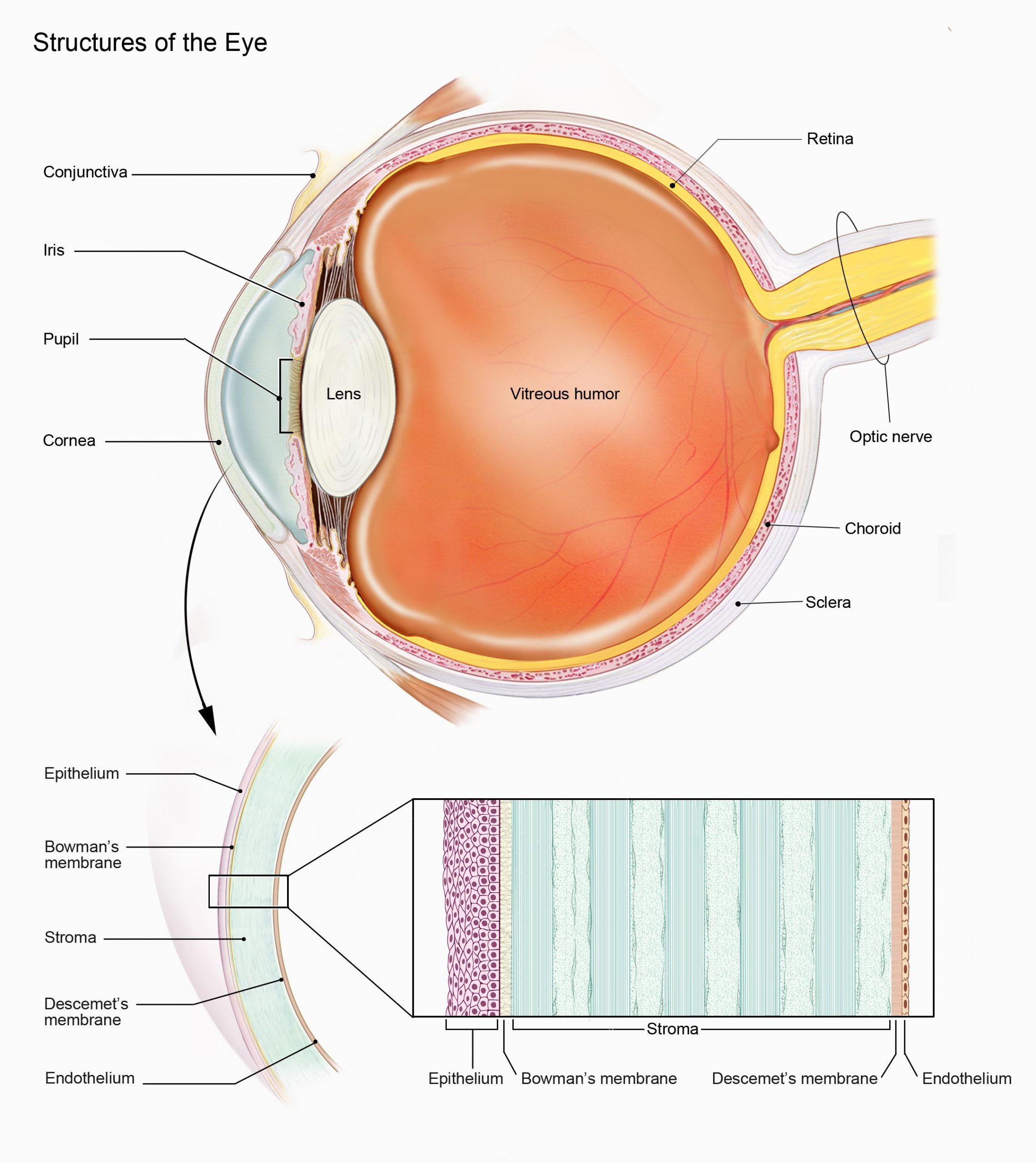
The images we see must be clear and sharp, and there are many components of the eye that enable this to happen. Firstly, the cornea, which is located on the front of the eye, is a transparent layer that allows light to pass through the eye and is mostly responsible for converging the light towards the retina, contributing around +42.00D of power to the optical system.
The crystalline lens is located within the eye, and this also plays a part in converging the light to focus on the retina, contributing around +18.00D of power. It is this lens that becomes less transparent when a cataract forms and is ultimately removed and replaced with an artificial lens.
If the power of the optical system is too strong, this indicates myopia, and a concave, diverging lens is needed to reduce the overall power. If the power of the optical system is too weak, this is hypermetropia, and a convex, converging lens is needed to increase the overall power.
The crystalline lens is attached to the ciliary muscle via zonular fibres, and it is this that allows the lens to alter its power to focus on near objects, a process known as accommodation. When we look at a close object, the ciliary muscle contracts, which allows the lens to bulge outwards and increase its overall power. This means the eye can adjust its focus.
The iris is the coloured part of the eye, and this contains two powerful muscles: the sphincter and the dilatator. The sphincter is responsible for reducing the pupil size, and the dilatator for increasing it. The main function of the iris is to alter the amount of light entering the eye, so it will reduce in size (constrict) when it is bright to reduce the amount of light entering the eye, and increase in size (dilate) at night, to allow more light through.
A clear fluid called the aqueous humour circulates around the anterior eye, and this pressurises the globe. Glaucoma is a condition when the pressure within the eye rises, and this can cause damage to the eye and ultimately sight loss. It is often symptomless, and for this reason anyone over 40 years of age with a family history of glaucoma is entitled to an NHS eye examination.
The other transparent fluid within the eye is the jelly like vitreous humour. This fills the posterior chamber of the eye and helps to keep the shape of the eye and supports the retina.
In order to see perfectly, all these components need to be in perfect balance, which illustrates how amazing our eyes are.
Sue Deal FBDO R is a practising dispensing optician, ABDO College examiner, senior tutor and supervisor for dispensing opticians. She is also a practice visitor and external moderator for ABDO.
OA Corner Part 1: What makes a good OA
OA Corner Part 2: Communications
OA Corner Part 3: A question of strategies
OA Corner Part 4: Understanding bifocals